Abstract
Between April 1998 and June 1999, eight palm vipers (Bothriechis marchi) were diagnosed with
a disease similar to inclusion body disease (IBD) of boid snakes.1,2 Signalments and clinical signs are
summarized in Table 1. Six palm vipers were captive bred and two palm vipers were wild caught. All palm vipers were
adults at time of death with a mean age of 8.6 yr. Three palm vipers were found dead with no premonitory clinical signs
and five palm vipers had one or more of the following clinical signs: anorexia, regurgitation, paresis, and dehydration.
All palm vipers had intracytoplasmic, round to oval, single to multiple, eosinophilic inclusion bodies in hepatocytes and
renal tubular epithelial cells. Inclusion bodies were distributed in other organs with varying frequency. The
distribution of inclusions in affected vipers is summarized in Table 2. Concurrent histologic lesions are summarized in
Table 3 and included urate nephrosis, septic thrombi, and hepatocellular degeneration. Ultrastructural features of the
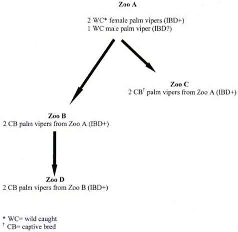
inclusions had features similar to inclusions in boid snakes with IBD.2 Figure 1 depicts a flow chart pedigree
analysis of the affected palm vipers. Based upon pedigree analysis, there appears to be a familial tendency for this
disease in this population of palm vipers.
Table 1. Breed, gender, age, and clinical signs of eight palm vipers (Bothriechis marchi)
with inclusion body disease.
|
Case number |
Gender |
Age (years)a |
Clinical signs |
|
1 |
Mb |
5 |
Regurgitation, anorexia |
|
2 |
Uc |
Adult |
Anorexia |
|
3 |
Fd |
13 |
Paresis, anorexia |
|
4 |
M |
Adult |
None |
|
5 |
F |
8 |
None |
|
6 |
M |
8 |
Anorexia |
|
7 |
F |
9 |
Dehydration, anorexia |
|
8 |
F |
Adult |
None |
a. Age at time of death.
b. Male
c. Undetermined
d. Female
Table 2. Distribution of inclusion bodies in eight palm vipers (Bothriechis marchi)
categorized according to frequency.
|
Location of ICIBa |
Cases |
Location of ICIB |
Cases |
|
Hepatocytes |
8 |
Glomerular mesangial cells |
4 |
|
Renal tubular epithelial cells |
8 |
Myenteric ganglia |
3 |
|
Respiratory epithelial cells |
6 |
Oviductal epithelial cells |
3 |
|
Biliary ductal epithelial cells |
6 |
Pancreatic acinar cells |
2 |
|
Gastric mucosal cells |
5 |
Esophageal mucosal cells |
3 |
|
Intestinal mucosal cells |
4 |
Epididymal epithelial cells |
2 |
|
Striated myofibers cells |
4 |
Thymic/splenic lymphoid cells |
2 |
|
Smooth myofibers |
5 |
Adrenal cells |
1 |
|
Thyroid follicular cells |
3 |
Pancreatic ductular cells |
1 |
a. ICIB= intracytoplasmic inclusion body.
Table 3. Concurrent histologic lesions in eight palm vipers (Bothriechis marchi) with
inclusion body disease.
|
Histologic lesion |
Cases |
Histologic lesion |
Cases |
|
Urate nephrosis |
5 |
Hepatitis |
2 |
|
Intravascular septic thrombi |
5 |
Gastroenteritis |
2 |
|
Hepatocellular degeneration |
5 |
Aortic mineralization |
2 |
|
Biliary ductal hyperplasia |
3 |
Interstitial pneumonia |
2 |
|
Granulomasa |
3 |
Hepatic melanosis |
2 |
a. Granulomas located in one or more of the following: skin, stomach, thyroid gland, intestine, lung, and
heart.
| Figure 1. | 
Flow chart depicting the distribution of the original wild caught palm vipers
(Bothriechis marchi) and the subsequent generations to different zoological parks. |
|
| |
Acknowledgments
We thank Dr. Suzan Murray (Fort Worth Zoo), Dr. Wynona Shellabarger and Dr. Tim Reichard (The
Toledo Zoo), Dr. Scott Gearhart and Dr. Gary West (San Antonio Zoo), Dr. Ann Duncan (Detroit Zoo), and David Heckard
(Abilene Zoo) for case submissions.
References
1. Axthelm MK.1985. Clinicopathologic and virologic observations of a probable viral
disease affecting boid snakes. Proc Am. Assoc. Zoo Vet. Pp. 108-109.
2. Schumacher J, ER Jacobson, BL Homer, JM Gaskin. 1994. Inclusion body disease in boid
snakes. J Zoo Wildl. Med. 25:511-524.