Abstracted from Infectious and Parasitic Diseases of Koi Copyright 1994, 1996, 1998 by Nicholas Saint-Erne, DVM
Definitions
 Disease--any deviation from the normal structure or function of
any part of the body
Disease--any deviation from the normal structure or function of
any part of the body
Pathogen--any disease producing microorganism (viruses,
bacteria, fungi)
Infection--invasion and multiplication of pathogenic
microorganisms in body tissues
Infestation--invasion of the body tissues by parasites
(protozoa, helminthes, arthropods)
Parasite--a plant or animal which lives upon or within another
living organism at whose expense it obtains some advantage
Host--a plant or animal that harbors or nourishes another
organism
Communicable--capable of being transmitted from one organism to
another (contagious)
Stress--the sum of all biological phenomena elicited in an
organism by adverse external influences
Environment--all the conditions, circumstances, and influences
surrounding and affecting the development of an organism
Clinical Sign--observable evidence of a disease state
Fishes--the plural of 'fish' when referring to more than one
species of fish.
Taxonomy of Koi
Kingdom--Animalia (animals)
Phylum--Chordata (chordates)
Subphylum--Vertebrata (vertebrates)
Superclass--Gnathostomata (jawed mouth)
Grade--Osteichthyes (bony fishes)"Teleostomi"
Class--Actinopterygii (spiny finned fishes)
Superorder--Ostariophysi (bone air bladder)
Order--Cypriniformes (carp shaped fishes)
Suborder--Cyprinoidae (carp-like fishes)
Family--Cyprinidae (carps and minnows)
Genus--Cyprinus
Species--carpio Linnaeus 1758
Variety--Japanese Colored Carp, or Koi, from "Nishiki Goi"
which is Japanese for "Brocaded Carp", a domestic bred variety of the common carp
Other closely related carp:
Common (wild) carp--Cyprinus carpio
Mirror carp--common carp with reduced number of scales
Leather carp--common carp with no scales
Crucian carp--Carassius carassius
Prussian carp (European)--Carassius auratus gibelio
Goldfish (Asian)--Carassius auratus auratus
Carp in the genus Cyprinus all have two pairs of barbels on the upper lip (maxilla). Carp in the genus Carassius do not have
barbels.
Introduction
Fish diseases can be very perplexing and frustrating for the koi pond keeper. So many things can affect the pond and make the fish
go belly up.
The most important aspect in maintaining fish health is proper water quality. This depends on such things as pH, temperature,
alkalinity, ammonia, nitrite, nitrate, oxygen concentration, population density, and biological, chemical and mechanical filtration systems. When all of
these parameters are within their proper ranges, the fish will have less stress and be more immune to disease.
Infectious and parasitic diseases occur more easily in fish stressed from improper environmental conditions. In the wild, fish may
harbor a variety of parasites without incurring problems. But, the artificial environments in ponds or aquaria are more likely to be abnormal, therefore
parasites can then cause serious diseases.
A combination of the environment, nutrition, genetics, and the presence of pathogens or parasites is involved in the development of
disease. Anything we can do to improve these will improve the health of the fish.
The following is a guide to the infectious, parasitic, nutritional, and environmental diseases of koi, with information on how to
diagnose, treat and prevent these problems.
Patient History
Prior to the actual examination of the fish, some information should be gathered. What abnormal signs does the fish exhibit? Which
fish are sick (all or a few or only one)? How long have they appeared to be ill? Are they still eating? What are they being fed? What type of filtration
system is used? When was it last cleaned? When was the last water change? How frequently are the water changes made? When was the water tested, and what
were the results?
Many times the answers to these questions will provide significant clues to the type of disease process affecting the fish.
Diagnostic Methods
A live moribund fish should be examined when possible, or a freshly dead specimen. If the fish has been dead for more than a few
hours, even if refrigerated, the chance of accurate diagnosis is diminished.
If the fish is alive, observe its respiration by watching its mouth movements and the motion of the operculum (gill cover). Note how
it positions itself in the water. Is it neutrally buoyant (normal) or does it float or sink? Does it list to one side, or float with its head down? Are
there lesions on the head, body or fins? Are the fins congested or eroded? Are any scales missing? Lift the operculum and examine the gills for
hemorrhage, discoloration, or necrosis.
After the initial physical examination, biopsy samples should be taken. Place the fish on a wet towel or chamois cloth, and wrap it to
keep it from jumping. Uncover sections of the fish as necessary to take biopsy samples. If Tricaine Methane Sulfonate (MS-222 anesthetic is available, use
this at 80-120 mg/L in fresh dechlorinated water to anesthetize the fish prior to the biopsy. Have another container of dechlorinated water without
anesthetic available in which to awaken the fish.
Using a blunt blade or spatula, or even a plastic microscope coverslip, gently scrape a small sample of mucus off the body of the
fish. Place this in a drop of water on a microscope slide and cover it with a coverslip. If there are skin or fin lesions, take a sample from the margin
of the lesion and prepare it the same way. Next snip a small section of the caudal or other fin rays using small sharp scissors, such as iris or suture
scissors. Also take a sample of one or two gill filament tips. Place these on a slide and prepare as with the skin scraping. Examine these samples
microscopically at 40 to 400X magnification.
A fresh fecal sample can be collected with a pipette or syringe from the container of water in which the fish arrived. Place this on a
microscope slide and compress it with a coverslip. Examine it for bacteria, protozoa, and helminth ova.
If the fish is dead upon presentation, obtain the same biopsy samples, but a larger portion of gill tissue should be examined. Then an
internal examination of the abdominal organs can be made. Collect tissue samples for bacterial cultures and sensitivities, histopathology, or microscopic
squash preparations, as appropriate.
Viral Diseases
Carp Pox
This disease is caused by Herpesvirus cyprini. It produces superficial milky-white to gray colored plaques on the skin and
fins. They extend 1-2 millimeters above the surface of the skin, and are usually smooth. Adjacent plaques may merge into larger raised areas. The lesions
are benign and eventually slough. The disease is not fatal but can produce scarring.
The virus is transmitted by contact of an affected fish with healthy fish. Overcrowding in the pond will increase the prevalence of
the disease. No treatments are completely effective against viruses, but application of a disinfectant solution on the lesions may be helpful.
Spring Viremia of Carp (Infectious Dropsy)
This European fish disease is caused by Rhabdovirus carpio. It produces a wide variety of signs, including a distended
ascitic abdomen (dropsy), inflamed and protruding vent, uncoordinated swimming, weak respiration, and hemorrhages on the gills and skin.
The virus is shed in the feces, and the gills are the site of virus entry and primary replication. Virus will spread in the blood to
the internal organs. Eggs can be infected at breeding. Fish lice and leeches can also carry the virus between fish.
The disease is more severe in colder water temperatures, hence the prevalence of signs in spring. Warming the pond water up to 68
Fahrenheit (20 Celsius) will protect the fish from the virus. Ultraviolet irradiation of the pond water will inactivate the virus and help prevent its
spread. Formalin added to the pond (25 mg/L) will also inactivate the virus. A vaccine can be made from formalin-inactivated virus and administered by
intraperitoneal injection. This will not help already infected fish, but will prevent infection in healthy fish. They should be vaccinated in the summer
or fall to prevent disease outbreak in the spring.
Bacterial Diseases
Bacterial Hemorrhagic Septicemia
This serious disease is caused by a number of species of bacteria from the Aeromonas genus. Most often identified is
Aeromonas hydrophila (A. Liquefaciens and A. punctata), but A. sobria, A. schuberti, A. veronii, A. saccharophila and A.
caviae may also be found. The organisms are motile, gram-negative, rod-shaped bacteria about 1 x 2.0-4.5 micrometers in size. They have a single polar
flagellum.
Aeromonas bacteria are almost always present in pond water, and even on the fish itself. It is an opportunistic pathogen that
proliferates when the fish is weakened or injured. Signs of infection include hemorrhages of the gills, skin and base of fins, loss of scales, and skin
ulcers. Eye swelling (exophthalmia) and abdominal swelling (dropsy) may also occur. Parasites such as fish lice cause skin wounds which then can become
infected with Aeromonas.
Treatment consists of antibiotic injections, antibiotics in the food or water, and topical applications of antimicrobial solutions
directly on the skin ulcers. Water quality improvement is indicated for all disease conditions.
Carp Erythrodermatitis (Furunculosis, Ulcer Disease)
Severe epizootics of this disease occur most frequently in the spring when the water temperature is rising (increased bacterial
growth) and the fish are stressed from over-wintering (decreased fish resistance). The signs are erythema (redness) on the skin and base of fins. Raised
skin lesions (furuncles) form which then ulcerate exposing the underlying muscles. Congestion and hemorrhage of the internal organs also occurs.
The causative organism is atypical Aeromonas salmonicida, a gram-negative, non-motile, short rod (1 x 2 micrometer) bacterium.
It occurs singly, in pairs, chains, or clumps. It is inactive in water temperatures below 44.6 Fahrenheit (17 Celsius). Disease outbreaks occur when water
temperatures rise above this in the spring. Treat as with other bacterial infections. Prevent osmotic stress due to skin lesions by adding salt to water
(0.1-0.3% solution).
Bacterial Fin and Gill Rot
This is usually caused by the gram-negative, short motile rod bacteria Pseudomonas fluorescens, but Aeromonas or other
species of bacteria can also be involved. Fin and tail erosions are the usual signs, but hemorrhages and skin ulcers sometimes occur. Injury to the fins
may result in this secondary bacterial infection.
Treat with antibiotics in food or water, antibiotic injections, and topical wound disinfection. Severely damaged fin margins can be
trimmed off with a scalpel or sharp scissors, and the fin topically disinfected. This may hasten healing and fin regeneration. Fin rot is commonly
associated with poor water quality.
Flexibacter columnaris (Cotton Disease)
This opportunistic pathogen causes reddened areas on the skin with cottony white growths, often mistaken for fungi. The lesions
occur mostly on the lips and fins, and across the top of the caudal peduncle. The bacterium is a long (10-12 microns), thin flexingly motile rod.
It will grow in water from 39-95 Fahrenheit (14-35 Celsius), but is more pathogenic in water above 64 F (118 C). On the surface of the
fish, large masses of this bacteria will align themselves into "haystack" formations. It occurs on previously injured tissues. Most healthy fish are
resistant to this infection.
Treat with antibiotics in the food, and with copper sulfate baths. The disease is worse at higher water temperatures so lower the
temperature if possible to slow bacterial growth. Other stresses (poor water quality, other diseases) make this organism more virulent (pathogenic).
Fungal Diseases
Saprolegnia, Achlya
These freshwater Oomycetes occur as external fungal infections on koi fish. They appear as white, cottony tufts on the skin or
gills, and also on fish eggs. Microscopic examination shows transparent non-septate branching hyphae. Fungal growth usually occurs on tissue that has been
previously damaged by trauma or other disease conditions. The fungus attacks unfertilized fish eggs, but once established will spread to the viable eggs.
Remove any white opaque eggs from the spawning mats if possible to prevent their decay.
Treatment consists of adding Formalin-Malachite Solution to the water, and using topical disinfectants on the affected skin. Fungus
proliferates in ponds with large amounts of organic debris (dead plants and leaves, dead fish, uneaten food), so detritus removal and water changes are
helpful.
Prevent fungal growth on egg masses with Methylene Blue (1 milligram per gallon of pond water daily for 3 days), or Hydrogen Peroxide
3% (1 milliliter per gallon of water).
| Differentiation of Saprolegnia and Achlya by zoosporangia and zoospores | 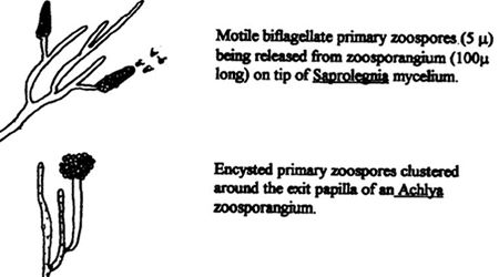
|
|
| |
Branchiomyces sanguinis (Gill Rot)
This fungus invades the blood vessels of the gills, obstructing the flow of blood. Infected gill filaments become brownish with
white or gray streaks. Microscopically it has branched non-septate hyphae.
High levels of unionized ammonia in the water increase the incidence of fungal gill rot due to gill epithelial cell hyperplasia. Death
can occur rapidly in affected fish, due to anoxia.
Treat with methylene blue and salt baths. Prevent by keeping the water cool and avoiding accumulations of organic debris.
Protozoal Parasites
Diagnosis of protozoal infestation is best done by examination of a live fish, or a freshly dead refrigerated fish. Frozen specimens
or fish dead for very long will not yield accurate samples of protozoa.
Affected fish should have a small area of skin scraped and the mucus examined under a microscope. A small section of a fin or of a
gill filament may also be clipped off for examination. A blood sample can be drawn and checked for blood parasites. Feces should be examined for
intestinal protozoa.
Spore Forming Protozoa (Sporozoa)
Eimeria, Goussia (Coccidiosis)
This is an intracellular protozoal parasite that causes inflammation of the intestines. Affected fish are thin and depressed. The
feces are often yellowish. Microscopic examination of the feces will reveal the coccidioid oocysts (20-40 microns). Each oocyst contains four sporocysts,
each of which contains two sporozoites.
Treat with tetracycline antibiotic in the food (250 mg per 100 grams of fish food). Feed the medicated food for 7-10 days.
Ciliated Protozoa (Ciliata)
Chilodonella
This ciliated protozoan attaches to the skin and gills causing respiratory distress, depression, fins clamped to sides, and
excessive mucus production which makes the skin look white or bluish. The gills may appear pale and covered with mucus. A smear taken from the mucus will
contain many of the organisms. The organism is notched at the posterior pole, giving it a heart-shaped appearance. It is 20-40 x 30-80 micrometers in
size.
It is an especially important parasite during the winter, when the fish are dormant. Chilodonella can reproduce in cold water
and prefers temperatures as low as 41-50 F (15-10 C). They reproduce by simple division (binary fission). Their numbers can increase when the fish is in a
weakened state from the cold water. Fission ceases at 68 F (120 C), and the parasite dies in warm water.
Treat with Formalin-Malachite Solution, copper sulfate, or a 2% salt solution for a 10-20 minute bath.
Epistylis, Heteropolaria
These ciliated parasites attach to the body, fins, and gills of fish by a long retractable stalk. They are filter feeders which
ingest organic debris and do not feed on the fish itself, but use it as a substrate for attachment. This can lead to secondary Aeromonas or
Saprolegnia infections at the attachment sites. Heavy infestation causes light colored cotton-like patches on the skin. Fish may scrape their sides
on rocks (flashing) because of irritation. They may also attach on fish eggs, causing them to appear fuzzy. Formalin-Malachite Solution will reduce the
growth of these parasites. Decreasing the organic load in the pond is also important.
Ichthyophthirius multifiliis (Ich)
These holotrichously ciliated parasites embed in the epithelium of the gills, fins and skin. They appear as small white spots on the
fish. Hyperplasia of the gill epithelium makes the gills look pale and results in hypoxia. Flashing is very frequently seen.
The embedded feeding stage (trophozoite, or trophont) grows in the epidermis for 3-30 days, depending on temperature. After feeding,
the trophont (up to 1 mm in size) breaks out of the epidermis leaving epithelial destruction. It drops from the fish to the bottom of the pond where it
encysts (tomont stage) and divides by binary fission within the cyst. Hundreds of free swimming tomites (20 x 50 micrometers) are released from the cyst
after 8-24 hours. These then search out a host fish on which to feed. They must attach to a host within 24-48 hours or they will die. The tomites secrete
hyaluronidase to allow penetration of the fish's epithelium. There it matures into the trophozoite feeding stage, completing the cycle.
The optimum water temperature for the ich parasite is 70-77 F (21-25 C), at which it will complete a full life cycle in 3-7 days. At
lower water temperatures it takes longer; as long as 5-6 weeks at 50 F (110 C). The free swimming tomites will not survive in water warmer than about
84-86 F (129-130 C). This can be useful in treating for this parasite. Formalin-Malachite Solution or copper sulfate will also kill the free swimming
tomite stage. Salt in the ponds at 0.1% (1 gram NaCl/liter water) will inhibit Ich infestations.
Click on the image to see a larger view.
1) Koi infested with Ichthyophthirius trophonts. 2) Trophonts (trophozoites)--embedded feeding stage: spherical cell completely
covered in cilia with a U-shaped macronucleus, up to 1 mm diameter. Grows in epidermis of fish until mature. 3) Trophont leaves host when mature and
attaches to plant, gravel, or other substrate. There it forms a cystic capsule (tomont stage) and begins mitotic cell division. 4) Mitotic division
continues for up to 10 times (210 = 1024 cells) producing hundreds up to a thousand new protozoa. 5) The tomont's cystic capsule ruptures and
the free-swimming ciliated tomites (theronts) are released. 6) Free swimming tomite will die if it does not find a host within 24-48 hours. 7) Tomite
penetrates and embeds in the epidermis of the skin or gills, where it matures as a trophont.
Trichodina Complex
Several genera of similar protozoa cause this disease. They are peritrichously ciliated, circular parasites that are flattened
ventrally (40-60 micron diameter). They live on the skin and gills where they damage tissue with their rotating denticular ring. Affected fish may produce
excessive mucus and develop a white cast to the skin. Small hemorrhages may appear on the skin and fins.
Formalin-Malachite Solution is effective in killing these parasites. Individually infested fish may be bathed in a 2% salt solution
(20 grams NaCl/liter water) for 10-20 minutes to remove these parasites.
Flagellated Protozoa (Mastigophora)
Hexamita (Octomitus), Spironucleus
These similar organisms infest the intestines of fish. In many cases they do not cause disease, but in large numbers they can cause
inappetence, weight loss, and even death. Stress from shipping, overcrowding, poor water quality, or malnutrition allows rapid reproduction of these
protozoa in the intestinal tract, leading to disease. Microscopic examination of feces will show highly motile protozoa with six anterior flagella and two
posterior somatic flagella. They are 4-8 x 10-12 microns in size.
Improved nutrition, fresh vitamin C (ascorbic acid) supplementation, and metronidazole in the food (250 mg/28 g food) or in the water
(250 mg/10 gal water) can be used for treatment.
Ichthyobodo necator (Costia necatrix)
This small (10x20 micrometer) flagellated protozoan attaches to the skin and gills by a hold-fast organelle. The parasite feeds on
the epithelial cells of the fish, resulting in sloughing of the epidermis. A bluish-white film may develop on the skin from excessive mucus production.
The fish will have respiratory difficulty and be depressed. Death occurs by asphyxiation due to damage of the gill epithelium.
It has two long flagella that project posteriorly from the body and cause the parasite to swim in a jerky spiral, which can be seen
microscopically on skin biopsy. It reproduces by binary fission and four flagella, two long and two short, may be seen prior to division. It spreads from
fish to fish by swimming, and it will encyst when conditions become unfavorable. It will survive in temperatures from 36-86 F (12-30 C), but its optimum
temperature for reproduction is 75-77 F (2425 C).
Treat with Formalin-Malachite Solution, or 2% salt bath for 10-20 minutes. This will kill the trophic (feeding) stage, but the
encysted stage may survive, so treatment should be repeated on a weekly basis as long as necessary.
Trypanosoma, Trypanoplasma, Cryptobia
These flagellated blood parasites live within the circulatory system of fish and can be found in the gills. They can cause
obstructions in the blood vessels of the gills. Signs include lethargy, weight loss, anemia and ascites. They are spread between fish by leeches.
Diagnosis is made by examining blood smears under a microscope. They appear as undulating flagellated cells moving rapidly among the red blood cells.
Collect the blood from the gills or from the ventral vein in the caudal peduncle.
Prevent the disease by controlling leeches.
Click on the image to see a larger view.
Cnidosporidia
Myxobolus koi
This myxosporidian parasite lives in the connective tissue of the gills of koi. The spores can obstruct blood flow in the gill
filaments, causing damage resembling bacterial gill disease. The mature spore (10 micrometers) consists of an oval bivalvulid shell around two uninucleate
sporoplasms and one polar capsule. The spores are released when the fish dies, and are ingested by an intermediate host (an invertebrate). The sporoplasms
are released, transforming into actinosporea. These leave the intermediate host and enter the fish host by penetration or by ingestion.
Formalin-Malachite Solution (25 mg Formalin and 0.10 mg Malachite Green per liter of water) may have some effect in preventing the
free-swimming actinosporean stage.
Helminth Parasites
Monogenean Trematodes (Flatworms)
These parasitic worms have a complete life cycle that involves only a single host.
Dactylogyrus (Gill Flukes)
These monogenean trematodes live on the tips of the gills and occasionally on the skin of fish. They cause gill filament hyperplasia
resulting in hypoxia. Signs include rapid respiratory movements, clamped fins, and flashing. Gill tip biopsy will reveal the flukes upon microscopic
examination. They have a four-pointed anterior end with four dark eyespots and a sucker disc. The posterior end (opisthohaptor) has one or two large pairs
of hooks and 12, 14, or 16 smaller peripheral hooklets.
Each fluke is hermaphroditic, having both a testes and an ovary, and releases a single large egg (40 microns) after mating with
another fluke. The egg has a hooked projection which may keep it attached to the fish, or to aquatic plants. The eggs hatch in 1-4 days. Newly hatched
flukes (oncomiracidia) are ciliated and swim to find a new host. They attach to the gills, or onto the body where they will then crawl to the gills. On
the gills, the larvae will mature in 3-6 days. The adult parasites often die in cold water in winter, but the eggs are capable of surviving the winter on
the bottom of the pond as low temperature will arrest their development.
Gyrodactylus (Skin Flukes)
These monogenean flukes occur mainly on the skin and fins, but occasionally are found on the gills. They are hermaphroditic and give
birth to live young, one at a time. An embryo with hooks is often visible microscopically within the adult. The young are parasitic immediately after
birth.
They cause localized hemorrhaging of the skin and excessive mucus production. They have two points on the anterior end and an anterior
sucker, but no eyespots. The opisthohaptor has one or two pairs of median hooks and eight pairs of marginal hooklets.
Treat flukes with Formalin-Malachite Solution, Trichlorfon, Praziquantel, or salt baths.
Digenean Trematodes
Fish serve as secondary intermediate hosts for these, which require multiple hosts (snails, fish, birds) to complete their life
cycle. The immature trematode is encysted in the tissue of the fish, and matures to adult when the fish is eaten by another animal. The encysted trematode
is a 1-4 mm black, white, or yellow "grub" seen in the muscles, internal organs, or eyes.
Eliminating snails from the pond will prevent transmission of the immature stage (cercaria) to the fish. Surgical excision of the
cysts is possible if they are near the surface of the fish's body. Intramuscular Praziquantel injections (25 mg/kg body weight, one time) may eliminate
the encysted metacercariae.
Sanguinacola (blood flukes) This digenean trematode uses fish as its final host. It lives in the blood vessels, heart, and the
gills. Its triangular shaped eggs can cause blockages of the circulatory system. The eggs hatch releasing miricidia which penetrate the gills and enter
the water in search of their intermediate host, a mollusc. After developing to the cercarial stage in the mollusc, they return to the fish and re-enter
the gills. There they reach sexual maturity and deposit eggs.
Gill damage occurs from the larval forms' penetrations, and from blood vessel blockages. Secondary bacterial infections can occur on
the necrotic tissues.
Eliminating snails from the pond will prevent the spread of this parasite. Affected fish can be treated with a Praziquantel injection
(25 mg/kg BW, intramuscularly).
Cestodes (Tapeworms)
Fish serve as primary hosts for tapeworms in their intestinal tracts. They also are secondary intermediate hosts for encysted
tapeworms (pleurocercoids) in their muscles and abdomen.
Caryophyllaeus fimbriceps
This tapeworm is parasitic to the intestines in koi. Its intermediate hosts are oligochaete annelids of the genera Tubilex and
Limnodrilus. Large numbers of tapeworms can cause intestinal inflammation and weight loss.
Treat with Praziquantel 0.50% added to the food. Avoid feeding live oligochaete worms to koi.
Nematodes (Roundworms)
Capillaria is found within the fish's intestines. Its attachment to the wall of the intestines can lead to areas of necrosis
and secondary bacterial infections. It may reduce growth rate and reproductive ability of the fish.
Diagnosis can be made from microscopic examination of a fecal sample. Ova with bipolar caps should be seen. Treat with Fenbendazole
0.25% in the food.
Leeches
These annelid worms are blood sucking parasites. They can be introduced into the pond with plants, rocks, snails, or on fish.
Leeches can cause anemia and can spread blood parasites between fish. After feeding they leave the host fish to breed. The hermaphroditic adults mate and
lay egg sacs in the pond substrate. Leeches have a mouth within an anterior sucking disc (oral sucker) and an anus in the posterior sucker.
Leeches should be picked off fish when seen, and the skin swabbed with a disinfectant. Trichlorfon (0.25 mg active Trichlorfon per
liter water) will kill leeches in the water.
Click on the image to see a larger view.
Crustacean Parasites
Lernaea cyprinacea (Anchor Worms)
These copepod crustacea have free swimming larval stages. Once mature, the adult male (170 x 780 micrometers) will mate with a
female and then die. The fertilized female develops an anchor shaped head which it embeds into the skin of the host fish.
This causes a reddened sore which may become secondarily infected with bacteria. The females develop two large egg sacs on their
external end, containing several hundred eggs each. The egg sacs are shed when the eggs are ready to hatch. The nauplii hatch from the eggs and break out
of the egg sac. The female will then develop new egg sacs. At the optimal temperature of 77 F (25 C), egg sacs are produced every two weeks. The mature
female may be up to 4 cm long.
Treat by gently extracting the parasite with forceps, being careful not to leave any of the anterior anchor behind. Treat the wound
with topical disinfectant. Trichlorfon organophosphate can be used to kill the free living juvenile stage. Lernea are also inhibited by 2% salt solutions
for short duration (10-20 minute) baths.
Ergasilus (Gill Maggots)
These parasitic copepods attach to fish's gill filaments with specialized claws formed from the second pair of antennae. They feed
on blood and body tissues. Only the female is parasitic. After they mate, the male dies and the female finds a host on which to feed while developing
eggs. Two large white egg sacs on the female's posterior resemble maggots. After the eggs hatch the juveniles are free living in the pond until they
mature and mate.
Treat with Trichlorfon (0.25 mg/L water).
Argulus foliaceus (Fish Lice)
These are oval, flattened Branchiurans with two prominent eyes, two sucking discs, and a stylet mouthpart. This stylet repeatedly
pierces the skin of the fish and releases toxic secretions. They feed on the tissue fluids released by this trauma. They are freely moving on the host and
swim well. Affected fish flash frequently. Skin ulcers from secondary bacterial or fungal infections are common on koi affected with Argulus.
The adult female fish louse lays a thin strip of eggs on submerged objects near the banks of the pond. The larvae hatch in 2-8 weeks,
depending on temperature. They immediately attach on fish to feed, maturing in 3-6 weeks. Adults can grow to 1 centimeter long. Reproduction ceases in
temperatures below 57 F (114 C).
Mosquito fish and bitterlings may eat immature forms of the parasite. For best control, treat the pond with Trichlorfon on a weekly
basis for 4-8 weeks.
Koi Pond Examination and Water Quality Analysis
Dr. Nicholas Saint-Erne
Date: _________
Location: ___________________________
Pond Size: _________
Filtration System: ___________________________
Fish Examination: ___________________________
Water Examination
|
Water quality |
|
Optimum range |
|
Temperature |
_________ |
65-75 F |
|
Oxygen content |
_________ |
6-9 mg/l |
|
pH |
_________ |
6.5-7.5 |
|
Alkalinity |
_________ |
25-250 mg/l |
|
Ammonia |
_________ |
0-0.01 mg/l |
|
Nitrite |
_________ |
0-0.01 mg/l |
|
Nitrate |
_________ |
0-20 mg/l |
|
Other recommendations: |
__________________ |
|
Recheck date: |
__________________ |
Disease Prevention Protocol
Quarantine new fish for 14-21 days
Monitor water quality frequently
Do partial water changes regularly
Keep filters clean and functioning properly
Feed a nutritionally balanced diet
Remove detritus from pond
Use proper hygiene (disinfection) on nets and equipment
Ultraviolet light will kill free swimming stages of many
parasites and microorganisms. Clean the light often to prevent reduction of irradiation capacity.
Activated carbon will adsorb chemicals from the water, so it
should be removed when medicating the pond. Filtration should continue in order to provide oxygenation.